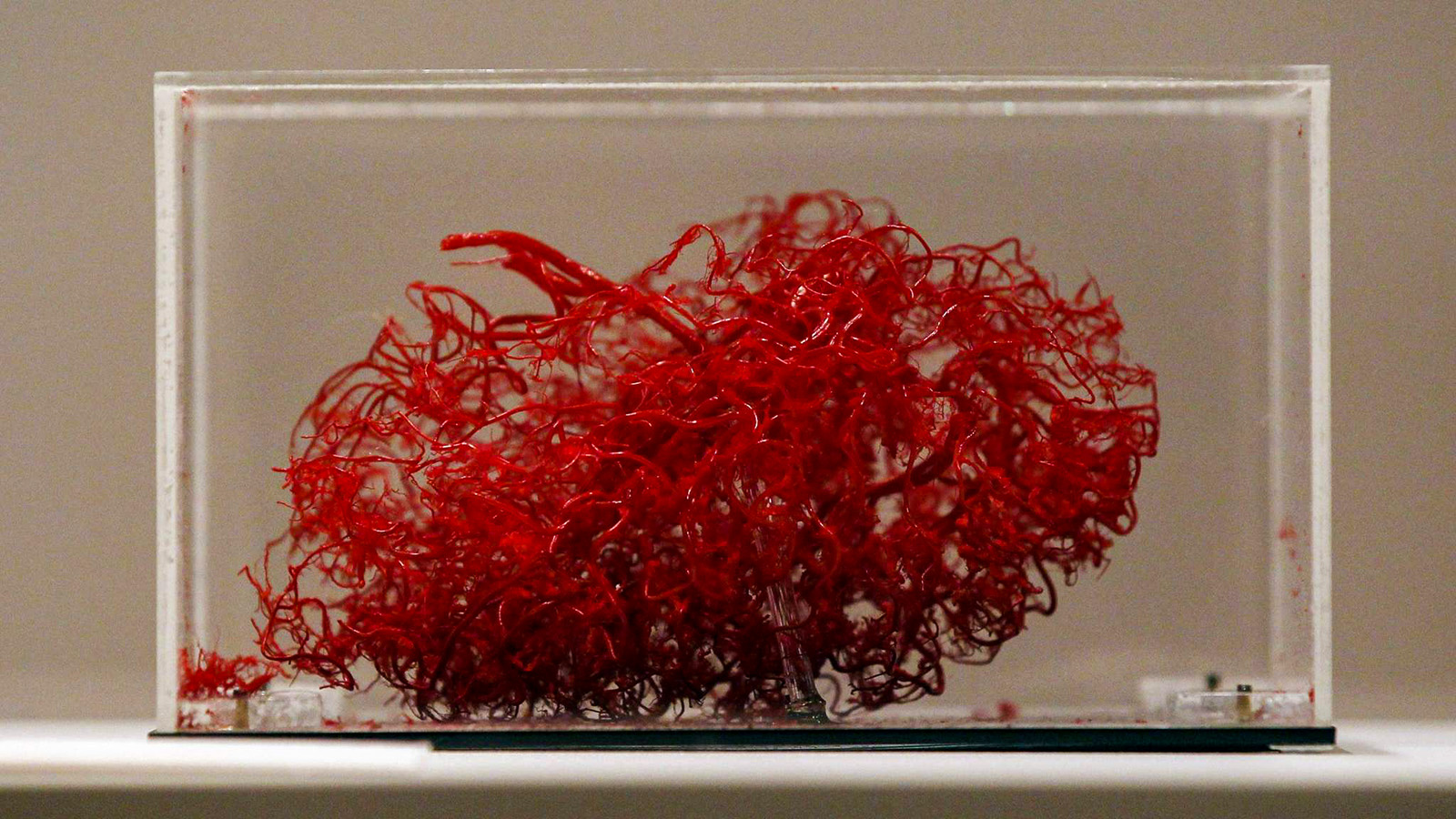
Bio-ink, bioreactors, organ moulds: this might sound like science fiction to those not familiar with three-dimensional tissue printing. We have learned about the technology promising to revolutionise healthcare in the University of Pécs János Szentágothai Research Centre, where researchers are working, among others, on printing the smallest bone in the human body: the stapes.
written by Miklós Stemler
One characteristic of incredibly expensive technologies is that they do not look expensive all that much – and this is exactly what we would think when looking at the tissue printing lab in the University of Pécs János Szentágothai Research Centre (UP JSR). Most of the equipment here is known from traditional cell growing laboratories, which is of course not a coincidence: on one hand, keeping cells alive is necessary for the tissue printing process, and on the other han, important events happen on the microscopic cell level, therefore the constantly developing and changing tools serve this miniature world.
An office printer with extras
This is true for the equipment we looked at, where average-looking laboratory equipment have price tags of a hundred million. As we learn from professor dr. Judit Pongrácz, head of the UP Pharmaceutical Biotechnology Department, one of their most important tool is a three-channel bioprinter, which means that it can print three different types of cells or any arbitrary combination of them. This does not mean much to a layman, but our guide gave a descriptive example to describe the process of bioprinting. “The simplest way is to imagine an office colour printer that has multiple cartridges for making colour: one is yellow, and the other is red. Instead of colour cartridges, we use various types of cells, this will create the ‘letters’. In contrast with a regular two-dimensional office printer, the bioprinter goes over the same letter multiple times, giving it the necessary thickness.”

Bioprinting has therefore two very important advantages over simple cell cultures: it can put multiple types of cells in an arbitrary combination in a three-dimensional structure, just like how living tissue and organs are constructed and functioning. This is the first step that is followed by many others, or is hoped to be followed by many others. The end points are numerous. The one that is the most exciting is a functioning, implantable human organ, but there are many problems to be solved by researchers until then – even when bio- and tissue printing are playing a large role in various therapy methods and research. Another endpoint is modelling organs and their diseases, providing opportunity for developing new medications and their combinations and for toxicology analysis.
The stem cell revolution
The foundation stone of bioprinting is the cell, of which many are needed, since a human organ is made up of billions of them. For a long time, reaching the critical cell number was the main obstacle: this was solved by a Nobel-winner discovery. The breakthrough in increasing cell numbers was made in 2006, remembers dr. Judit Pongrácz. This is when a Japanese researcher, Jamanaka Sinja successfully transformed adult mouse cells into stem cells; cells that can be ‘reprogrammed’ into other types of cells. The researcher repeated the process successfully on human cells a year later. The discovery received a Nobel-prize in 2012, and it is incredibly important for regenerative medicine aiming to make use of the healing properties of the human body – it provides an opportunity to use the so-called inducted pluripotent stem cells for reparing damaged tissue. This method allows a reasonably quick and easy way to grow a large number of cells, and then used them for better study of our organs and tissues, thereby developing new methods of healing.

Using inducted stem cells means that therapies like replacing dead heart tissue after a coronary could be made possible and commonplace (these have only been tested on animals). All of this is of course not as simple as it sounds: there are many trials and experiments to be made until the truly efficient cell transplantation solutions are found for human use. Researchers and clinicians had an important revelation when it turned out that the behaviour of the implanted cells is influenced by the cell environment (meaning that cells implanted into a diseased environment will become diseased themselves), and this was the base of the development of bioprinting, where tissue reproduction happens in a complex form, aiming for transplanting tissue or entire organs instead of single cells.
All of this resulted in even more problems to solve. Reproducing an organs does not only need the right type of cells, but also extensive knowledge of it: what the structure of the organ looks like, its morphology during function. And organs do not exist separately from each other but as a part of the body’s ecosystem, therefore knowing and recreating this environment is also necessary for the longevity of the bioprinted tissue. This means that before we can start printing any kind of organ, we need to analyse the organ and its environment with the most modern imaging methods.
The magic wand effect
After all that is done, the actual printing can start, with the goal of recreating the three-dimensional structure of the tissue. There are working methods here: but we are still very far from creating complex organs like the heart – but there are examples for cartilages and bones. “If a bones is damaged in an accident and the only option is replacement, it is possible now to mature bones is various laboratories after creating the necessary support structure and implanting the right type of cells” – explains dr. Judit Pongrácz.
The word ‘mature’ is used on purpose, since aside from spatial organisation, the fourth dimension – time – is also essential: printed organ initiations need to develop and grow just like their traditionally grown counterparts. For this an environment similar to that of a living system is needed, this is what we call bioreactors, where the organ initiations can develop. Aside from creating the necessary environment, the young tissue needs to be trained as well in order to ensure it will function properly in the future and to turn into real, functional cartilage or bone that can handle the weight load. Similarly, we can only create functional lungs in an environment that can simulate real circumstances, since the movement of the lung tissue and its contact with air is what defines the development of a healthy, functional organ.
This all means a lot of experimenting and temporary failures. “We have to figure out step by step, what kinds of cells we need to use, what environment they thrive in, how nutrients get to each cells, what interactions happen. if we put everything together correctly and set the most important factors correctly, we will see that our lab-grown cells to what they should be doing, just like we have waved a magic wand – it shows the reward after the long time spend experimenting.”
Bio-inks – we are no longer just dreaming about them
The most serious challenge those dealing with bioprinting face is that of creating three-dimensional structures, and encourages printed cells to form these. There have been multiple breakthroughs in this area in the past few years, and now there are so-called base matrixes available that can help promote the development of the printed tissues. These base matrixes are especially important in creating soft tissue. While in bone printing, outlining the correct shape and size of the printed organ is done by using pre-printed and absorbable base structures, cells of soft tissue can be shaped and encouraged to form tissue formations with the base matrixes. Soft matrixes became especially popular, since the hard base structure also raises problems, since these need to be absorbed without posing danger to the host body after the tissue is ready and implanted.
Technologies and materials that can be used to encourage independent cell growth to create structure by themselves were created to replace the pre-printed frames – this are the so-called bio-inks. “While traditional frames have the cells added on them, bio-ink means we can surround the developing tissue, creating a permeable barrier that protects the cells and provides the ideal environment, but is still permeable by nutrients and oxygen” – explains dr. Judit Biri-Bovári, a member of the research group.

Bio-inks provide much artistic freedom, adds dr. Judit Pongrácz, which is important because tissue printing is widely used for pharmaceutical testing, where individually created tissue allows testing the effects of new substances as widely as possible. The first step for this was provided by inducted stem cells, which made creating and analysing disease models from a patient’s own diseased cells possible, and bioprinting allows the highly accurate reproduction of the environment in a human body.
This is mostly important in pre-clinical trials, where possible harsh side effects can be filtered out in an environment modelling the human body with bioprinted complex tissues – this will make clinical stage trials involving humans a lot safer. Moreover, if the efficacy of a component underperforms, it can be detected at this stage already, which is a big time and money saver. All of these tests can be conducted on dozens of thousands of three-dimensional tissue samples with the continuous analysation of data, and therefore problems caused by genetic differences can also be detected.
Toxicology and efficiency studies of medicine and medicine-candidate agents are an important part of the research of dr. Judit Pongrácz. “One of the important areas is the testing of medicine to be used on rare diseases. Getting to know the mechanisms of the disease and then analysing the effects of already licenced medications on printed tissue is exciting and provides opportunities that give a lot of hope in therapies” – explains the head of the research.
From the smallest bone to the circulatory system
Although we are still separated from bioprinted, not rejected livers or hearts by many problems, bioprinting already has multiple clinical use cases. Many laboratories have reached success in creating ear or knee cartilage, which can make replacing and restoring damaged tissue possible.
Another big leap was made in the field of printing bones, researchers in Pécs are working on a project like this with the doctors of the Department of Otorhinolaryngology, including dr. Péter Bakó and dr. Máté Kocsis, and dr. Péter Maróti from the 3D Centre. They are working on the smallest bone in the body, the stapes in the ear, which is a challenge due to its small size. it is only 3.3x2.8 millimetres, and its unique shape makes printing difficult, needing true teamwork. “We have not mentioned yet that we are a part of a larger group. Experts and radiologists well-versed in the structure and function of a given organ make the best possible imaging of the printable organ possible, and that’s where we come in, creating a liveable tissue from that” – describes the process dr. Judit Pongrácz.
Aside from the bone printing clinical cooperation, researchers in Pécs are also dealing with soft tissue and the connecting circulatory system. According to researchers, the beauty of the area is that more and more details come to light about the functions and behaviour of tissues during their printing, or sometimes even unexpected interactions. Their ‘feedback’ can help making steps forward in getting to know the complicated system of the human body.

This is what attracts more students towards bioprinting as well, like a member of the research Alexandra Nagy biotechnology master’s student, who is working on the project differentiating stem cells into fat cells. “For me this field is very exciting because it is directly connected to getting to know medicine targets, the testing of developed medications, and correcting mistakes appearing in tissues, which all helps us learn a lot. Tissue printing raises many questions, all of which we would like an answer to – and then the answers lead to even more questions” – she says.
This excitement is what drives researchers worldwide, like dr. Judit Pongrácz who has been working with three-dimensional tissues for 26 years, and her colleagues. They want to realise the old dream: to exploit the regeneration skills of living tissue to use it for healing and lengthening life.
Photos:
Lajos KALMÁR
